
What is a Navicular Stress Fracture?
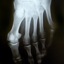
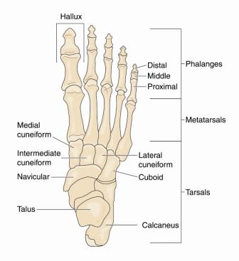
A navicular stress fracture is a term for a mild and incomplete fracture/break to the navicular bone which is located on the inside edge of the mid foot just below the ankle. The foot itself is made of three sections of different bones. The bones at the back of the foot under the ankle joint are known as the tarsal bones which include the calcaneus (which forms your heel), the talus (which forms part of the ankle), navicular, cuboid, and the lateral, intermediate and medial cuneiforms. The next section is the mid foot consisting of the five long metatarsal bones, numbered in the sense that your big toe is first and your little toe is your fifth. The metatarsals connect at the back to the cuneiforms, navicular and cuboid bones and at the front to the toes. The toes are the last section consisting of five bones called phalanges which like the fingers are individually divided into three parts (distal, middle and proximal) and the big toe is referred to as the hallux. The navicular bone plays an important role in the biomechanics of the foot. It contributes to and supports the structure of the foot arch thus affecting balance and supporting weight-bearing, it also provides the attachment point the tibialis posterior muscle (an important shin muscle).
What is the difference between a Navicular Fracture and a Navicular Stress Fracture?
A navicular stress fracture is similar to a navicular fracture only the fracture is incomplete and more like a crack than a break. Stress fractures are generally caused by an increase in repetitive stress placed upon the bone, hence the name stress fracture. However a full fracture is normally the result of a high impact injury or direct trauma to the foot.
Why and when does a Navicular Stress Fracture happen?
Navicular stress fractures are most common in the young people who participate in athletic sports. The stress fracture normally occurs as the result of a gradual build up in tension and micro-trauma from the connecting tibialis posterior muscle. When the tibialis posterior tightens or is overused typically from repetitive sports, in particular running and dancing, this increases the stress placed upon the navicular bone. This repetitive pressure normally elicits a stress reaction in the bone which can progress with continued pressure into a stress fracture. Wearing inappropriate and unsupportive footwear and training on hard unforgiving surfaces can both be factors responsible for causing a navicular stress fracture. Just as important a factor to consider is the biomechanics of your foot. Many cases of stress fractures in the foot are the result of flat fleet when the arches of your foot have fallen and in some cases when arches are too high. Poor foot posture, changes the way in which you walk and in turn increase the stress through the lower leg muscles.
What to do if a Navicular Stress Fracture is suspected after trauma?
If you suspect you have sustained a navicular stress fracture then it is important that you seek medical attention. If any type of fracture is suspected after trauma then you must immediately call for medical help or attend your local Accident & Emergency. Full fractures are considered a medical emergency due to the possible risk of damage to the surrounding arteries and nerves.
What does a Navicular Stress Fracture feel like?
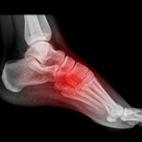
A navicular stress fracture causes a gradual onset of a dull ache around the fracture site on the inside of the foot. This pain can radiate down into the sole of the foot, up into the ankle and occasionally into the heel. The pain levels will feel quite low to begin with but will most likely be aggravated by sporting activities that include impact through the foot, for example running, football and dancing. The stress fracture may be accompanied by a small amount of swelling and bruising and in severe cases the pain levels will be aggravated by most ankle and foot movements. This makes weight bearing and walking very painful. The initial intense pain will ease with rest but once the healing process starts there will be a dull, continuous ache from the swelling and healing process that will last weeks to months. During the healing process the crack in the navicular bone is trying to knit together and re-unite. This stage can be quite painful and it is very important to rest and avoid weight bearing on the foot to ensure the navicular heals completely. The tibialis posterior muscle may be tight and contracted and may create stiffness and muscle pain around the shin and ankle areas.
How will a Navicular Stress Fracture be diagnosed?
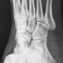
Your GP or Therapist will be able to diagnose you by both listening to your history and examining you. A full examination will be done to rule out any additional injuries or complications. Your doctor will most likely order an X-ray of the foot to confirm the diagnosis and identify the location and the extent of the stress fracture. Occasionally, if the extent of the fracture isn’t fully visible on an X-ray, an MRI or a CT scan will be ordered as this will give a more detailed view of the fractured site. Doctors may also use an MRI scan to assess whether the pain felt after trauma is coming from injury to the leg muscles, ligaments or joint capsule and not indeed from a fracture. The occurrence of a navicular stress fracture or full fracture from very mild trauma may prompt doctors to investigate for any underlying bone weakening conditions.
What treatment options are available for a Navicular Stress Fracture?
The treatment options and healing times for navicular stress fractures vary depending on the position and severity of the fracture and also on the complications from any additional injuries. Most cases of navicular stress fractures heal within three months and do not need surgical intervention, however it is occasionally necessary to surgically stabilise multiple fractures or complete breaks. Patients will initially have to rest and avoid aggravating activities. In most cases you may be initially required to avoid weight bearing and to use crutches, a boot splint or a cast to reinforce and stabilise the fracture for the first six weeks until it is well healed. The doctor will most likely prescribe anti-inflammatory medication to reduce any swelling and painkillers for pain relief. Patients will be referred to a physiotherapist who will provide exercises to strengthen the ankle, foot muscles and ligaments to aid the healing process. It is vital that during the healing process the patient still moves as much as possible to reduce the risk of a DVT. Fluctuating pain levels will be experienced throughout the healing process however most of the discomfort towards the end of the healing process comes from stiffness levels caused by long term immobility. Rehabilitation exercises as well as massage and some manipulative therapies like osteopathy or chiropractic can aid in recovering foot and ankle flexibility once the fracture site has healed.
If your GP has mentioned or if in fact you feel that there is some element of poor foot posture that has predisposed you to this injury then it is very important that you see a podiatrist who can fully examine your feet and determine whether you need orthotics for your shoes. The podiatrist should also be able to provide you with advice on more appropriate and supportive training shoes. The failure to remedy these problems may result in a prolonged recovery or recurring stress fractures.